

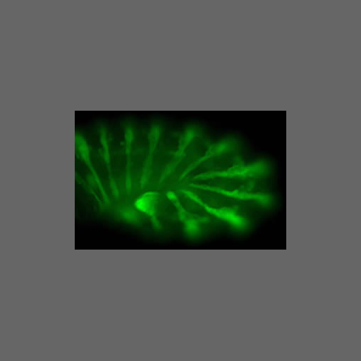


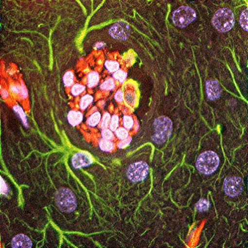
Top: 9L tumor cells (rat gliosarcoma) (at 200X magnification) growing and spreading along brain capillaries of athymic nude rats. Sample shows expression of GFAP (green fluorescence of reactive glial cells) and vimentin (orange-reddish fluorescence of 9L cells). Nuclei are bluish-purple fluorescent (glia and neurons) and intense pink (9L cells). .
Bottom: Blind deconvolution of the red, green, and blue components removes the 'haze' without producing artifacting. (Image from Dr. Alexandru C. Stan from the Hannover Medical School in Hannover, Germany.)


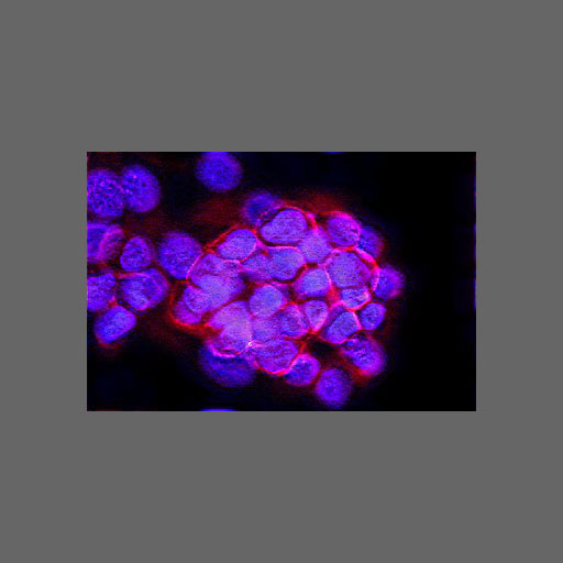
Top: This is an image of cells taken by a confocal laser scanning microscope. This image was also provided by Dr. Stan.
Bottom: The deconvolution shows structures within the cells that could not be clearly be seen before processing.


This is an image of a live cell also taken by a confocal laser scanning microscope. The restoration was limited by the bit depth of the image. For greater resolution, we would love to combine ImPASS with con-focal imaging. If you are in this field, ask us!


Top: The image is an interferogram of water drops containing some motion blur that was generated by imaging thin liquid films on surfaces. Since the fluid within the duration of the exposure, the images are often slightly blurry. The objective is to remove the blur such that there is more contrast in the fringes.
Bottom: The motion blur has been diminished and more fringes appear to the right. Some artifacting is seen in objects that are not in the same focal plane as the majority of the image. (Image courtesy of Prof. Joel Plowsky, Chemical Engineering, RPI.


This is an image (top) is of microbes taken from the base of Mount St. Helens, as seen through an optical microscope. Fine structures and increased constrast appear in the restoration.


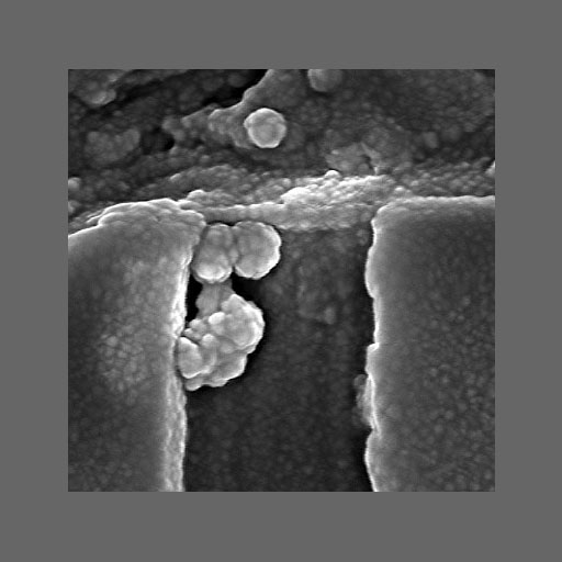
Top: An example of non-optical imaging. Scanning Electronic Microscope image of gold sputtered cross-section integrated circuit at effectively 600,000x magnification.
Bottom: Blind deconvolution removes the haziness, increase contrast, and reveals structures that could not be seen before. (Image from Dr. William E. Vanderlinde formerly from the Laboratory for Physical Sciences, College Park, Maryland, USA.)